Lutetium-177 is the workhorse of today’s radioligand therapy: it is the radionuclide in Lutathera and Pluvicto. Terbium-161 is the challenger. It has almost the same half-life and chemistry, and a very similar beta spectrum, but each decay also releases a shower of low-energy conversion and Auger electrons. Most of those electrons travel only nanometres to a few tens of micrometres, so they dump their energy in or right next to the cell that took up the drug. In 2025 the VIOLET trial reported the first-in-human results of a terbium-161 PSMA therapy, ¹⁶¹Tb-PSMA-I&T [1].
How much does that extra electron shower actually add? In 2020 Alcocer-Ávila and colleagues at the University of Bordeaux answered that at the scale of single cells [2]. With their in-house track-structure code CELLDOSE, they calculated that terbium-161 delivers 2.6 to 3.6 times the dose of lutetium-177 to the nucleus of an isolated tumour cell, for the same total electron energy released.
Results from one code are a single data point, so we rebuilt the whole study with a different, independent tool: Geant4-DNA, the track-structure extension of the open-source Geant4 toolkit [3]–[8]. We ran it on a laptop, an Apple MacBook Pro with an M5 Pro chip. This article reports what we found: the results, a trap in Geant4’s terbium-161 data that every user should know about, and what the computation cost.
The question: what do low-energy electrons add?
Both radionuclides emit beta particles with a mean energy around 130–155 keV. In water, those electrons travel up to a couple of millimetres, far more than the 14 µm width of a typical tumour cell in this model. So from a single labelled cell, most of the beta energy escapes. That suits a bulky tumour, where neighbouring cells catch the escaping energy (the “cross-fire” effect), but it is wasteful against one isolated cell or a tiny cluster.
Terbium-161’s extra emissions change the balance. Per decay, the reference decay data (ICRP Publication 107 [9]) list:
| Electron energy per decay (keV) | Lutetium-177 | Terbium-161 |
|---|---|---|
| Beta particles | 133.3 | 154.3 |
| Conversion electrons (CE) | 13.52 | 39.28 |
| Auger electrons (AE) | 1.13 | 8.94 |
| Total electrons | 147.9 | 202.5 |
| Geant4, as simulated here (all electrons) | 147.9 | 202.0 |
Terbium-161 releases about 3.3 times as much energy as conversion and Auger electrons (48 vs 15 keV per decay), and about eight times as much as Auger electrons alone. Counting particles, our Geant4 runs gave about 14.7 electrons per terbium-161 decay against 2.4 for lutetium-177. Most of these are slow electrons with very short ranges.
The paper modelled exactly this situation:
- Cell: a 14 µm sphere of water (1,436 µm³) with a concentric 10 µm nucleus. The nucleus fills about 36% of the cell, on the large side, as in many cancer cells.
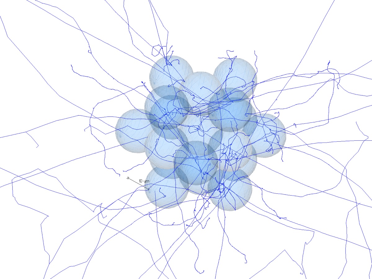
- Micrometastasis: a central cell with 6 first neighbours touching its faces and 12 second neighbours on the edges of a cube, 19 cells in all.
- Four source locations: the radionuclide spread uniformly on the cell surface, in the cytoplasm, in the nucleus, or through the whole cell.
- Fair comparison: every case was normalised to the same electron energy released per cell, 1 MeV per µm³ (1,436 MeV). The question is therefore “where does the energy go?”, not “which nuclide emits more?”.
- What was scored: the absorbed dose to the cell nucleus (where the DNA is), and for the cluster the fraction that comes from a cell’s own decays (the self-dose).
How we rebuilt it
We wrote a Geant4 application of about 1,200 lines of C++ that mirrors the paper’s model:
- Geometry: identical dimensions. Cells and nuclei are nested water spheres; the cluster is a simple cubic lattice of touching cells.
- Electron physics: Geant4-DNA (model set “option 2”) transports every electron collision by collision, down to 7.4 eV, the same cut-off CELLDOSE uses. It runs inside a water sphere that encloses the cells plus a 10 µm margin. Beyond that margin, a standard condensed-history model takes over. Our tests showed this hybrid changes the nucleus dose by about 1% and makes the cluster runs three times faster.
- Decay: Geant4’s own radioactive-decay module decays each terbium-161 or lutetium-177 nucleus at rest. It generates the beta particle, the conversion electrons and the full Auger cascade. As in the paper, photons are not tracked (their energy mostly escapes a cell anyway), but their energy is recorded.
- Statistics: 10⁵ decays per case (the paper used 10⁶). That gives 0.2–1.1% statistical uncertainty for the single cell and 1.4–2.6% for the cluster’s central cell.
- Software: Geant4 11.4.3 with its standard data libraries, built from source on macOS. The whole build took under three minutes on this machine.
Results: the single cell
The pattern is the same in both codes. The closer the radionuclide sits to the DNA, the higher the nucleus dose: 5.6 times higher for lutetium-177 and 7.7 times higher for terbium-161, comparing an intranuclear source with a surface source. Terbium-161 always wins. Its advantage grows as the source moves inward, because its slow electrons deliver most of their energy within a few micrometres of the decay.
Results: the micrometastasis
In the 19-cell cluster, every cell carries the same activity, so each nucleus also receives cross-fire from its neighbours.
| Source | ¹⁷⁷Lu, Geant4-DNA | ¹⁷⁷Lu, paper | ¹⁶¹Tb, Geant4-DNA | ¹⁶¹Tb, paper | ¹⁶¹Tb ÷ ¹⁷⁷Lu (ours / paper) |
|---|---|---|---|---|---|
| Cell surface | 6.88 Gy (30%) | 7.2 Gy (26%) | 14.19 Gy (32%) | 15.1 Gy (33%) | 2.06 / 2.1 |
| Cytoplasm | 7.63 Gy (36%) | 8.3 Gy (36%) | 16.93 Gy (48%) | 17.9 Gy (46%) | 2.22 / 2.2 |
| Whole cell* | 10.33 Gy (53%) | 11.0 Gy (53%) | 27.95 Gy (69%) | 29.1 Gy (67%) | 2.71 / 2.6 |
| Nucleus | 15.03 Gy (68%) | 15.7 Gy (68%) | 47.17 Gy (81%) | 47.8 Gy (81%) | 3.14 / 3.0 |
Dose to the nucleus of the central cell; the self-dose fraction is in brackets. *The whole-cell row was derived from the nucleus and cytoplasm runs: a whole-cell source is a volume-weighted mix of the two (0.364 × nucleus + 0.636 × cytoplasm). In the single cell, this shortcut matched a direct simulation to within 1%.
The neighbouring cells also agree. All 12 first- and second-neighbour doses are within 1–5% of the paper, and their self-dose fractions are within about one percentage point. For surface and cytoplasmic sources, cross-fire raises the dose to the central nucleus 2.7–3.7-fold for lutetium-177 but only 2.1–2.9-fold for terbium-161. That narrows terbium-161’s advantage to about 2× in the cluster, but does not remove it.
Of the 20 published self-dose fractions, 17 agree to within about one percentage point. The largest difference is lutetium-177 on the cell surface in the central cell: 30% in ours vs 26% in the paper. That value has the largest statistical error in our set. The paper’s figure also breaks a trend that holds everywhere else: for a surface source, the self-dose fraction rises from the centre outwards, from 30 to 32 to 40% in our results, and 26 to 32 to 40% in the paper.
How close is close?
All 28 values sit 1–8% below the paper. The shortfall shows no trend with nuclide, source location or cell position, and its spread is barely larger than the statistical noise. A uniform offset like this is the signature of a systematic difference between the codes, not of a modelling error. We tested three candidate causes:
- Decay data: ruled out. Geant4’s electron energy per decay matches ICRP 107 to within 0.3% for both nuclides, including the split between beta and conversion-plus-Auger energy. The normalisation uses the simulated energy in any case.
- Geometry and far-field transport: ruled out. Widening the track-structure region from 10 to 60 µm, or using track structure everywhere, moved the dose by at most about 1%. And the cluster reproduces the paper’s self-dose fractions, which are very sensitive to the geometry.
- Electron cross sections in water: the remaining explanation. CELLDOSE and Geant4-DNA use different physics models for how slow electrons ionise and scatter in water. Switching Geant4-DNA alone from model set “option 2” to “option 4” shifted the dose by 1.2%. Differences of a few per cent between independent track-structure codes are therefore plausible.
None of this touches the paper’s conclusions. Terbium-161 delivers about 2.6–3.6 times the nucleus dose of lutetium-177 in a single cell and about 2–3 times in a micrometastasis, and cross-fire dominates in the cluster for surface and cytoplasmic sources.
A trap for anyone simulating terbium-161 in Geant4
Our first terbium-161 results came out about 7% above the paper, while lutetium-177 matched. The cause was in Geant4’s decay data, not in our code.
Geant4’s decay file for terbium-161 labels each of its main beta branches as “first-forbidden”, a class of nuclear transition whose spectrum can differ from the standard (“allowed”) shape. For that label, Geant4 applies a single, generic shape correction. A comment in its own source code says the correction was fitted to bismuth-210 data and is “not valid for other 1st forbidden nuclei”. The result is a softer beta spectrum: a mean energy of 138.5 keV instead of the 154.3 keV in ICRP 107, which treats these branches as allowed. Lower-energy betas stay in the cell more readily, so the nucleus dose goes up.
The fix is small. We copied Geant4’s short terbium-161 decay file and removed the “firstForbidden” label from its six non-unique first-forbidden branches. A rare (0.01%) unique first-forbidden branch keeps its correct shape. Geant4 loads the edited file at run time with one command. The beta energy then becomes 154.8 keV and the total electron energy 202.0 keV per decay, matching ICRP 107. Lutetium-177’s file carries the label only on a negligible 0.016% branch and needs no change.
What it cost, and how to make it faster
All production runs used every core of the laptop: 18 threads on an Apple M5 Pro (6 performance cores, 12 efficiency cores, 64 GB memory). Memory was never a constraint, staying below 1 GB.
| Case | Time per decay | Time per run (10⁵ decays) |
|---|---|---|
| Lutetium-177, single cell | 4.4–4.6 ms | about 7.5 min |
| Terbium-161, single cell | 9.7–9.8 ms | about 16 min |
| Lutetium-177, 19-cell cluster | 8.7–8.8 ms | about 15 min |
| Terbium-161, 19-cell cluster | 15.7–15.8 ms | about 26 min |
Terbium-161 costs about twice as much as lutetium-177 because each decay produces about six times as many electrons, and track-structure physics follows every one of them collision by collision. The cluster costs about twice the single cell because the track-structure region is much larger. In total, the 14 production runs took 3.6 hours of computing; the whole project, including validation and sensitivity runs, took 4.2 hours. From the first “can this Mac run Geant4?” check to finished results took about six hours of elapsed time.
Measured this way, the full production set is worth about 40 performance-core-hours. The paper’s 10⁶ decays per case would be ten times that, about 36 hours on the laptop. The ways to cut that, in order of payoff:
- Don’t simulate what you can derive. A whole-cell source is a weighted sum of nucleus and cytoplasm sources (checked to 1%), and the neighbour doses come free with each cluster run. That removes 6 of the paper’s 16 configurations.
- Don’t over-sample. At 10⁵ decays the single-cell uncertainties are already 0.2–1.1%. Only the cluster’s central cell benefits from more statistics.
- Split by random seed, not just by thread. The job is embarrassingly parallel: independent runs with different seeds add up exactly. That means a workstation, a cluster or a fleet of cloud instances can share the work with no code changes.
- Use bigger x86 or Arm servers. At list prices, a 64-vCPU cloud compute instance costs roughly US$2.5–3.5 per hour on demand, and much less as spot capacity. Our rough estimate puts the paper’s full 10⁶-decay set at around 10 hours on one such machine, or 2–3 hours across four in parallel, for a total of a few tens of dollars. These are planning figures, not quotes; check current prices before budgeting.
- Package it in a container. A batch-only Geant4 image with its data libraries (about 2.5 GB) runs one macro with one seed per container, on any Linux host or cloud batch service.
What doesn’t help: GPUs (Geant4 and Geant4-DNA transport runs on CPUs), more memory, or more processes than physical cores.
Other lessons from the reproduction
Three more problems cost real time or would have produced wrong numbers silently:
- A messenger collision. Geant4’s
G4GenericMessengeridentifies commands by the last word of their path. Two commands ending in the same word, such as/cell/source/nameand/cell/output/name, cause one to be silently ignored. Our first runs simulated the wrong nuclide as a result. - Histogram units. In
G4AnalysisManager,CreateH1(..., 0., 600., "keV")reads the limits in Geant4’s internal unit (MeV) and uses the unit string only for display. Every entry landed in the first bin. The limits must be written as600.*keV. The doses were not affected, because they come from separate per-event sums. - Unit conversion in the analysis. The first analysis script applied the MeV-to-joule conversion twice and reported a dose of 10¹³ Gy. It was caught because the C++ application computes the dose independently. Keep two independent paths to every headline number.
What this means
An independent code, an independent implementation and independent decay data all reproduce the 2020 result to within a few per cent. That makes the dosimetric case for terbium-161 against small-volume disease more robust. At the cellular scale, its conversion and Auger electrons roughly triple the dose to the nucleus of a lone tumour cell, compared with lutetium-177.
The limits are the paper’s own. The model treats cells as uniform water spheres with the radionuclide evenly spread. Absorbed dose is not the same as biological effect. And getting a radiopharmaceutical into the nucleus, where terbium-161’s advantage is largest, is a hard drug-design problem. Clinical trials such as VIOLET [1], not simulations, will settle whether the physical advantage becomes a clinical one.
The practical lesson for dosimetry groups is simpler: if you model terbium-161 in Geant4, check the beta spectrum first.
References
- Buteau JP, Kostos L, Jackson PA, et al. First-in-human results of terbium-161 [161Tb]Tb-PSMA-I&T dual beta–Auger radioligand therapy in patients with metastatic castration-resistant prostate cancer (VIOLET): a single-centre, single-arm, phase 1/2 study. Lancet Oncol. 2025;26(8):1009–1017. doi:10.1016/S1470-2045(25)00332-8
- Alcocer-Ávila ME, Ferreira A, Quinto MA, Morgat C, Hindié E, Champion C. Radiation doses from 161Tb and 177Lu in single tumour cells and micrometastases. EJNMMI Phys. 2020;7:33. doi:10.1186/s40658-020-00301-2
- Incerti S, Baldacchino G, Bernal M, et al. The Geant4-DNA project. Int J Model Simul Sci Comput. 2010;1(2):157–178. doi:10.1142/S1793962310000122
- Bernal MA, Bordage MC, Brown JMC, et al. Track structure modeling in liquid water: a review of the Geant4-DNA very low energy extension of the Geant4 Monte Carlo simulation toolkit. Phys Med. 2015;31(8):861–874. doi:10.1016/j.ejmp.2015.10.087
- Incerti S, Kyriakou I, Bernal MA, et al. Geant4-DNA example applications for track structure simulations in liquid water: a report from the Geant4-DNA Project. Med Phys. 2018;45(8):e722–e739. doi:10.1002/mp.13048
- Kyriakou I, Sakata D, Tran HN, et al. Review of the Geant4-DNA simulation toolkit for radiobiological applications at the cellular and DNA level. Cancers. 2022;14(1):35. doi:10.3390/cancers14010035
- Agostinelli S, Allison J, Amako K, et al. Geant4 — a simulation toolkit. Nucl Instrum Methods Phys Res A. 2003;506(3):250–303. doi:10.1016/S0168-9002(03)01368-8
- Allison J, Amako K, Apostolakis J, et al. Recent developments in Geant4. Nucl Instrum Methods Phys Res A. 2016;835:186–225. doi:10.1016/j.nima.2016.06.125
- ICRP. Nuclear decay data for dosimetric calculations. ICRP Publication 107. Ann ICRP. 2008;38(3). icrp.org